Picture this. A patient walks in and says, "It hurts when I bite down on something hard, but only sometimes, and only on this one spot… maybe." No swelling. No visible decay. The X-ray looks completely normal. Your professor is watching. What do you do?
If your stomach just did a little flip reading that good. That instinct is correct. This is one of the few scenarios in dentistry where the "textbook normal" tooth is actually hiding something, and it has a name that intimidates clinicians more than it should be allowed to: Cracked Tooth Syndrome (CTS).
This isn't a topic we're covering because it sounds impressive on an exam. We're covering it because CTS is genuinely one of the most misdiagnosed conditions in general dental practice, and the research backs that up loudly.
Why This Condition Gets Everyone, Not Just Students
Here's the part that should make every dental student feel a little better about their own confusion: even experienced practitioners struggle with this. A clinical review on NCBI's StatPearls describes CTS as a condition marked by an unknown-depth fracture plane running through the tooth's structure one that can cause anything from occasional biting discomfort to full pulp involvement or root exposure, and its ambiguous nature demands a systematic diagnostic approach even from seasoned clinicians (StatPearls, NIH, 2024).
That phrase — "unknown depth fracture plane" is the whole problem in a nutshell. You genuinely cannot always tell how deep the crack goes just by looking, and how deep it goes determines everything about prognosis and treatment.
A published literature review on CTS puts it plainly: diagnosis is rarely straightforward, since the signs and symptoms often mimic other dental conditions, yet catching CTS early is critical to managing it successfully. The same review notes that even highly experienced practitioners find the diagnosis genuinely difficult, largely because associated symptoms vary so much from one patient to the next.
So if you've ever sat in clinic second-guessing yourself over a "maybe crack," join the club. It's a big club, and it includes people with decades of experience.
The Tooth That's Usually the Culprit
Interestingly, CTS isn't evenly distributed across the mouth. Reviews on the topic point to the second lower (mandibular) molar as the tooth most frequently affected, largely because of premature occlusal contact with malocclusion and deep or extensive restorations (including older amalgam fillings placed for retention) cited as leading causes of these fissures. Multiple sources agree that mandibular molars, in general, are the teeth most commonly affected by CTS.
So when a patient points vaguely at their lower back molar and says "it's this side, I think," your radar should already be up.
Why Your Diagnostic Tools Aren't as Reliable as You Think
This is the part dental schools sometimes gloss over: not all diagnostic tests are created equal, and some of the "classic" ones aren't nearly as accurate as you'd hope.
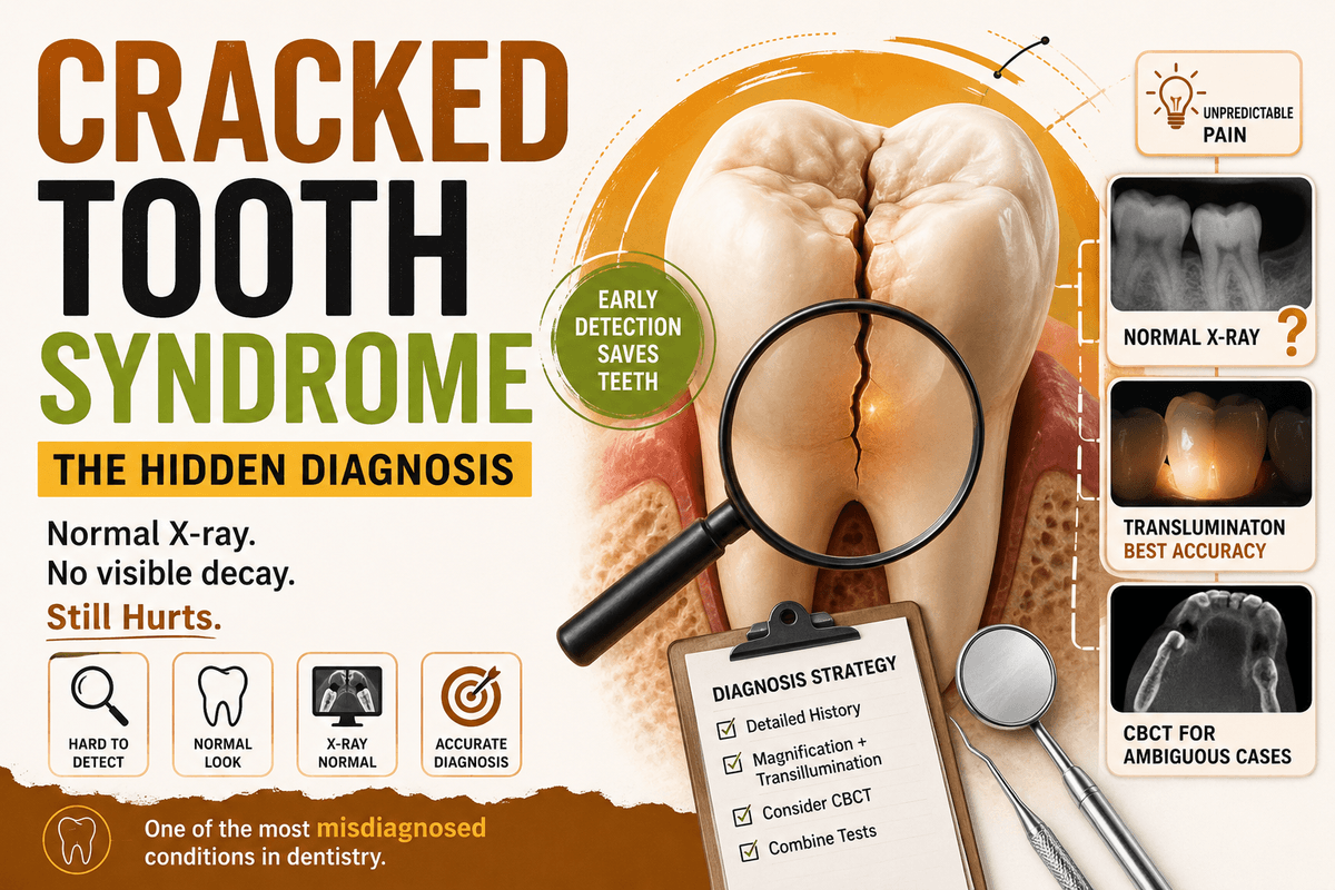
A 2025 diagnostic accuracy study compared four commonly used visual tests macrophotography, surgical microscope, transillumination, and DIAGNOcam against micro-CT as the gold standard. The results are worth sitting with. Transillumination came out on top for overall accuracy (65.3%) and sensitivity (68.8%) in diagnosing cracks. Macrophotography and high-magnification microscopy had the strongest specificity, at 92.9%, and high-magnification microscopy alone delivered the best positive predictive value, at 96.7%. Meanwhile, low-magnification microscopy trailed the pack with an accuracy of just 52.2% (Clinical and Experimental Dental Research, 2025).
Read that again: even your best single tool for spotting a crack only catches it correctly about two-thirds of the time. That's not a knock on you as a future clinician — it's a fact about the limits of the tools themselves, and it's exactly why combining tests matters so much.
On imaging, a mini-review from Frontiers in Oral Health adds an important caveat: while radiographs and cross-sectional imaging remain the most useful imaging methods overall, MRI and ultrasonography have been explored for crack identification but don't reliably pinpoint the lesion's exact location. Even laser-based and thermographic approaches often fall short of providing enough information for a confident CTS diagnosis on their own. The same body of research notes that cone beam CT has become genuinely indispensable in select, more ambiguous cases but that doesn't make it a magic bullet either. It's one more tool in a multi-test approach.
What's Actually Happening Inside the Tooth
If you want to understand why CTS causes such unpredictable pain, look at the histology. A study published via The Journal of the American Dental Association archives examined extracted cracked and worn teeth and found something striking: cracks were detected microscopically in every single specimen studied, and all of them were colonized by bacterial biofilms. Bacteria had invaded the dentinal tubules in these teeth, especially where the crack ran perpendicular into the dentin. In many specimens, the crack extended all the way to the pulp, triggering reactions ranging from mild inflammation to complete pulpal necrosis and symptoms tended to appear in most cases where the pulp itself was affected.
This is the biological reason CTS pain is so inconsistent: the crack itself becomes a highway for bacteria, and the tooth's reaction depends entirely on how far that highway has been built.
Where the Field Is Headed
Diagnosis-by-eyeball has an expiration date. Researchers are actively working on AI-assisted crack detection to reduce the guesswork. A 2022 paper reviewing imaging modalities for cracked tooth diagnosis noted that despite years of clinical and pre-clinical development, diagnosing a cracked tooth especially in its early stages remains a genuine challenge. That's exactly why image-processing and AI-based analysis are being explored as a next step. It's not replacing clinical judgment any time soon, but it's a clear sign of how seriously the profession takes this diagnostic gap.
The Practical Takeaway for Students
You are not going to nail CTS diagnosis with one test, one look, or one confident guess. The research is unanimous on this point across every review referenced above. Build the habit now, in school, of using a layered approach:
Take a real history timing, type of pain, specific triggers (biting vs. temperature sensitivity)
Use magnification and transillumination together, not either/or
Don't dismiss a normal-looking radiograph CTS often doesn't show up on standard films
Escalate to CBCT when the clinical picture and patient-reported symptoms don't add up
Remember that a "well-restored" or seemingly intact tooth can still be the cracked one some datasets have found intact or minimally restored teeth showing a higher incidence of cracks than heavily restored ones
Get comfortable saying "I'm not sure yet" to a patient. It's more honest and ultimately more useful than a false negative that turns into an emergency root canal six months later.
FAQs
Q: Is Cracked Tooth Syndrome the same as a visibly broken tooth? No. CTS specifically refers to an incomplete fracture that isn't always visible to the naked eye, unlike an obvious chipped or broken tooth. That's exactly why it's so easy to miss.
Q: Why doesn't a normal X-ray rule out CTS? Because most cracks run in a plane that doesn't show up well on a 2D periapical radiograph the crack has to be oriented just right relative to the X-ray beam to be visible, which is rare. This is part of why cone beam CT has become an indispensable tool for select, ambiguous cases.
Q: What's the single best diagnostic test for CTS? There isn't one "best" test that's the whole point. Current evidence shows transillumination has the strongest overall accuracy and sensitivity among common visual tests, while high-magnification microscopy offers the best specificity and positive predictive value. Combining tools consistently outperforms relying on a single method.
Q: Which teeth should raise my suspicion first? Mandibular (lower) molars, especially second molars, based on multiple reviews linking them to occlusal prematurities and malocclusion-driven cracking.
Q: Does clinical experience guarantee better crack detection? Not as much as you'd expect. Diagnostic accuracy studies have found only moderate improvements tied to experience level, reinforcing that this is a genuinely difficult diagnosis regardless of how many years someone has practiced.
Try goodoff